部分应用文献
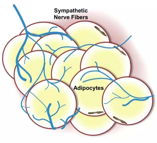
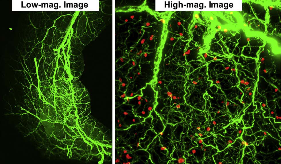
1) Dense Intra-adipose Sympathetic Arborizations Are Essential for Cold-Induced Beiging of Mouse White Adipose Tissue. Jiang H, Ding X, Cao Y, Wang H, Zeng W. Cell Metab. 2017 Oct
2) CUBIC pathology: three-dimensional imaging for pathological diagnosis. Nojima S, Susaki EA, Yoshida K, Takemoto H, Tsujimura N, Iijima S, Takachi K, Nakahara Y, Tahara S, Ohshima K, Kurashige M, Hori Y, Wada N, Ikeda JI, Kumanogoh A, Morii E, Ueda HR. Sci Rep. 2017 Aug
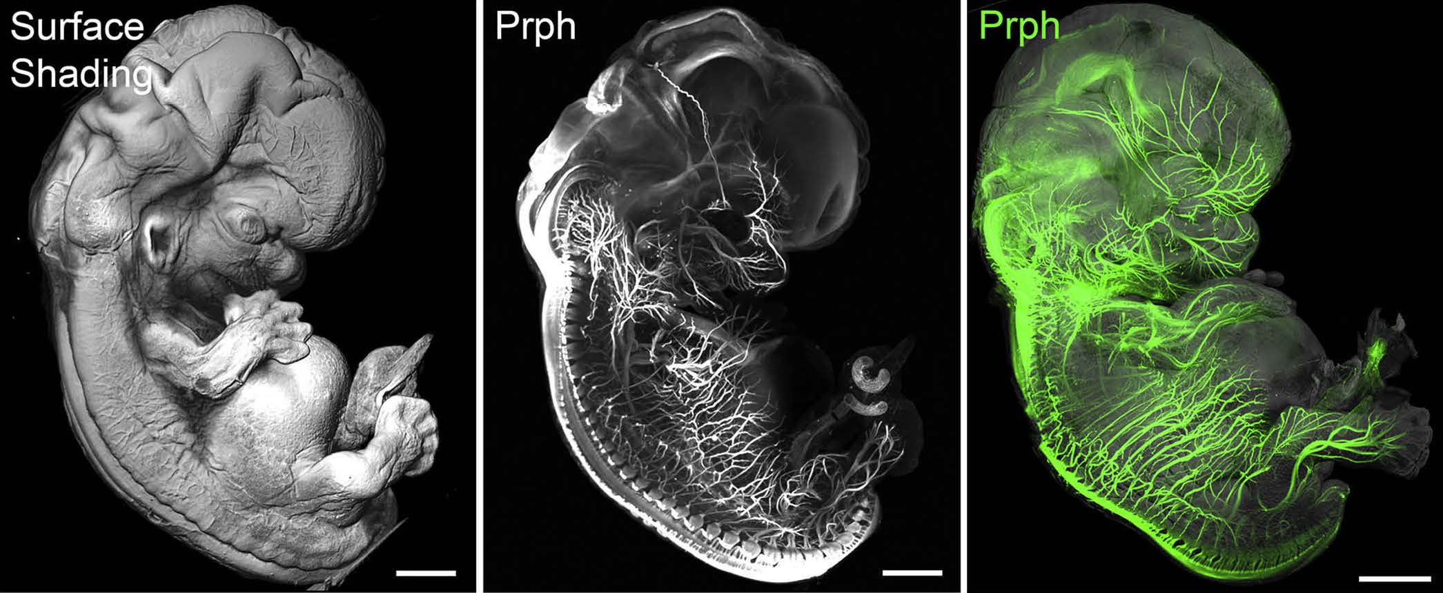
3) Tridimensional Visualization and Analysis of Early Human Development. Belle M, Godefroy D, Couly G, Malone SA, Collier F, Giacobini P, Chédotal A. Cell. 2017 Mar
4) Intra-islet lesions and lobular variations in β-cell mass expansion in ob/ob mice revealed by 3D imaging of intact pancreas. Parween S, Kostromina E, Nord C, Eriksson M, Lindström P, Ahlgren U. Sci Rep. 2016 Oct
5) Shrinkage-mediated imaging of entire organs and organisms using uDISCO. Pan C, Cai R, uacquarelli FP, Ghasemigharagoz A, Lourbopoulos A, Matryba P, Plesnila N, Dichgans M, Hellal F,Ertürk A. Nat Methods. 2016 Aug
6) Fully Automated Evaluation of Total Glomerular Number and Capillary Tuft Size in Nephritic Kidneys UsingLightsheet Microscopy. Klingberg A, Hasenberg A, Ludwig-Portugall I, Medyukhina A, Männ L, Brenzel A, Engel DR, Figge MT, Kurts C,Gunzer M. J Am Soc Nephrol. 2016 Aug
7) Three-Dimensional Study of Alzheimers Disease Hallmarks Using the iDISCO Clearing Method. Liebmann T, Renier N, Bettayeb K, Greengard P, Tessier-Lavigne M, Flajolet M. Cell Rep. 2016 Jul
8) Ultramicroscopy as a novel tool to unravel the tropism of AAV gene therapy vectors in the brain. Alves S, Bode J, Bemelmans AP, von Kalle C, Cartier N, Tews B. Sci Rep. 2016 Jun
9) Mapping of Brain Activity by Automated Volume Analysis of Immediate Early Genes. Renier N, Adams EL, Kirst C, Wu Z, Azevedo R, Kohl J, Autry AE, Kadiri L, Umadevi Venkataraju K, Zhou Y, Wang VX, Tang CY, Olsen O, Dulac C, Osten P, Tessier-Lavigne M. Cell. 2016 Jun
10) Wiring and Molecular Features of Prefrontal Ensembles Representing Distinct Experiences. Ye L, Allen WE, Thompson KR, Tian , Hsueh B, Ramakrishnan C, Wang AC, Jennings JH, Adhikari A, HalpernCH, Witten IB, Barth AL, Luo L, McNab JA, Deisseroth K Cell. 2016 Jun
11) Correlated magnetic resonance imaging and ultramicroscopy (MR-UM) is a tool kit to assess the dynamics of glioma angiogenesis. Breckwoldt MO, Bode J, Kurz FT, Hoffmann A, Ochs K, Ott M, Deumelandt K, Krüwel T, Schwarz D, Fischer M,Helluy X, Milford D, Kirschbaum K, Solecki G, Chiblak S, Abdollahi A, Winkler F, Wick W, Platten M, Heiland S,Bendszus M, Tews B. Elife. 2016 Feb
12) Advanced CUBIC protocols for whole-brain and whole-body clearing and imaging. Susaki EA, Tainaka K, Perrin D, Yukinaga H, Kuno A, Ueda HR. Nat Protoc. 2015 Nov
13) Combined 3DISCO clearing method, retrograde tracer and ultramicroscopy to map corneal neurons in a whole adult mouse trigeminal ganglion. Launay PS, Godefroy D, Khabou H, Rostene W, Sahel JA, Baudouin C, Melik Parsadaniantz S, Reaux-Le Goazigo A. Exp Eye Res. 2015 Oct
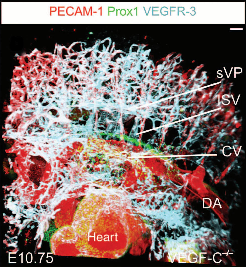
14) Podoplanin and CLEC-2 drive cerebrovascular patterning and integrity during development. Lowe KL, Finney BA, Deppermann C, Hägerling R, Gazit SL, Frampton J, Buckley C, Camerer E, Nieswandt B,Kiefer F, Watson SP. Blood. 2015 Jun
15) iDISCO: A Simple, Rapid Method to Immunolabel Large Tissue Samples for Volume Imaging. Renier N, Wu Z, Simon DJ, Yang J, Ariel P, Tessier-Lavigne M. Cell. 2014 Nov
16) Whole-body imaging with single-cell resolution by tissue decolorization. Tainaka K, Kubota SI, Suyama TQ, Susaki EA, Perrin D, Ukai-Tadenuma M, Ukai H, Ueda HR. Cell. 2014 Nov
17) A Simple Method for 3D Analysis of Immunolabeled Axonal Tracts in a Transparent Nervous System. Belle M, Godefroy D, Dominici C, Heitz-Marchaland C, Zelina P, Hellal F,Bradke F, Chédotal A Cell Rep. 2014 Nov
18) Fusing VE-cadherin to α-catenin impairs fetal liver hematopoiesis and lymph but not blood vessel formation. Dartsch N, Schulte D, Hägerling R, Kiefer F, Vestweber D. Mol Cell Biol. 2014 May 9) Multispectral fluorescence ultramicroscopy: three-dimensional visualization and automatic quantification of tumor morphology, drug penetration, and antiangiogenic treatment response. Dobosz M, Ntziachristos V, Scheuer W, Strobel S Neoplasia. 2014 Jan


